


Brain Disease Diagnosis
Medical image labeling services can support brain imaging research by annotating relevant anatomical regions and specified abnormalities in scan datasets. These labels help prepare training data for research models focused on brain tumors and other brain conditions.
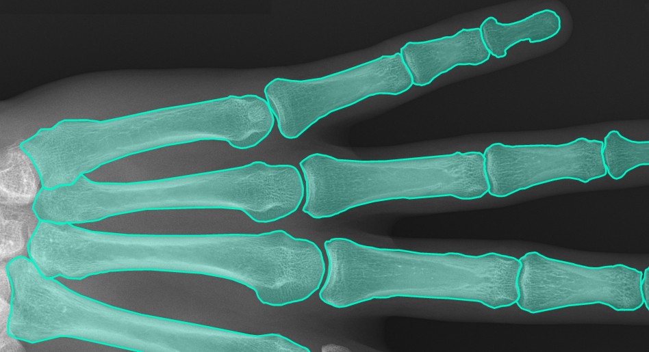
X- Ray Analysis
X-ray annotation services for medical AI label relevant structures and findings in radiographic images according to defined annotation guidelines. Potential annotation categories include:
- Bone fractures
- Specified tumors or abnormal findings
- Lung fluid-related findings
- Osteoporosis-related features
- Arthritis-related features
- Relevant blood vessel abnormalities, where visible and appropriate to the imaging modality.

Microscopic-Cell-Analysis
Medical image segmentation services can include contour-based annotation of microscopic cell images. Depending on the project, datasets may cover:
- Fibroblasts
- Lung cancer cells
- Liver cells
- Eye Cells
- Brain cells
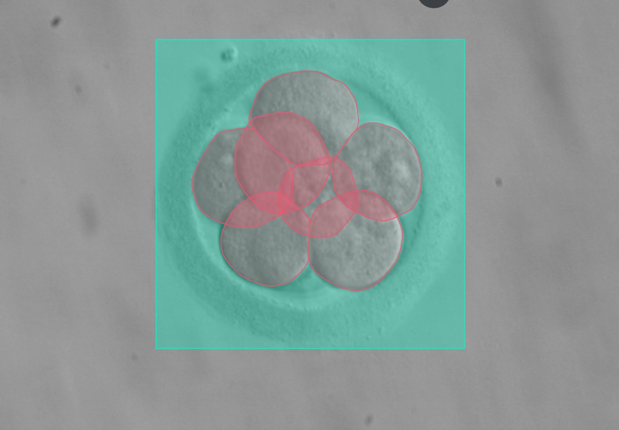
Embryo Cell Analysis
Embryo image annotation involves labeling specified structures in microscopy images, such as the zona pellucida and relevant cell regions. These datasets can support the research and development of computer vision models for assisted reproductive technology applications.
Eye Cell Analysis
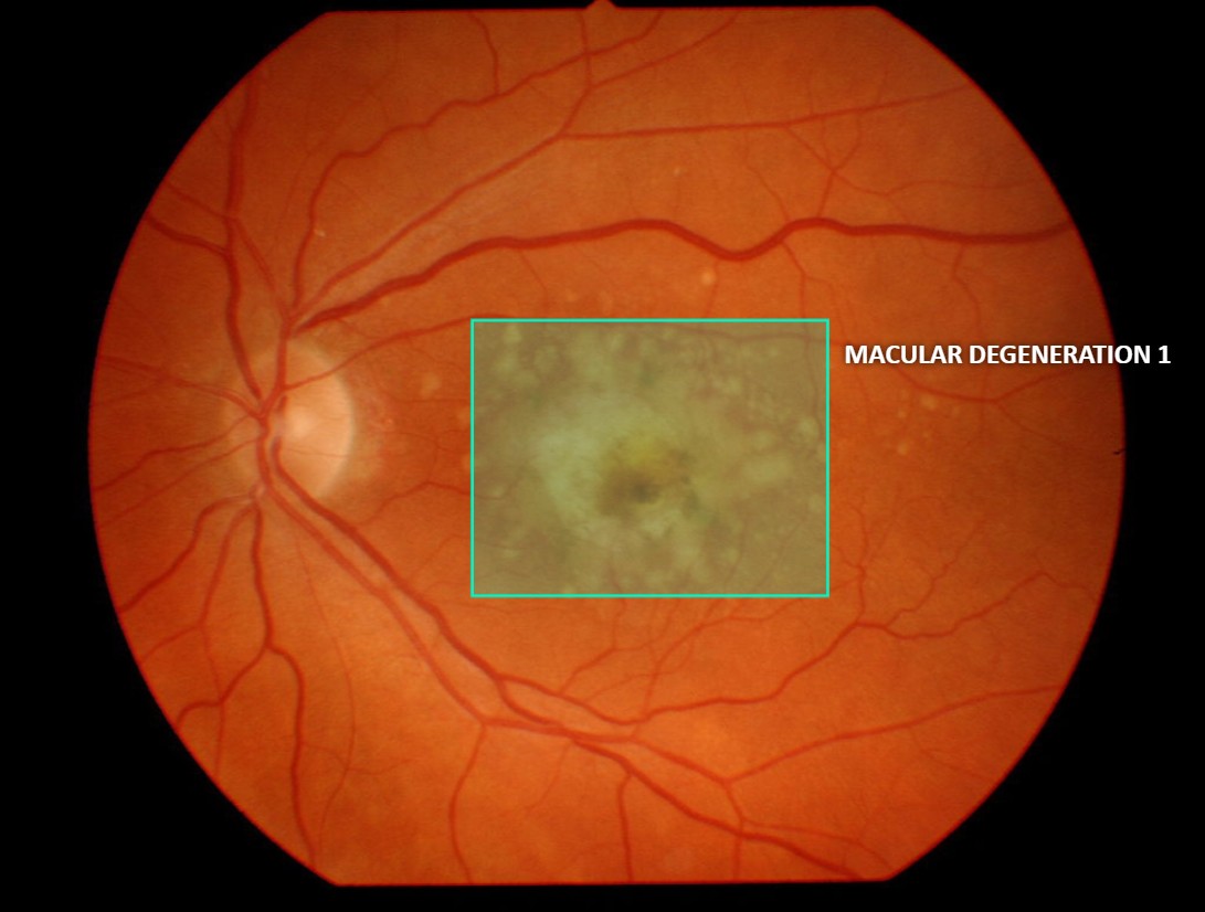
Retinal image annotation helps prepare structured datasets for computer vision research involving eye imaging. Based on project-specific requirements, annotations may cover features associated with conditions such as:
- Macular degeneration
- Diabetic Retinopathy
- Glaucoma
- Brain cells
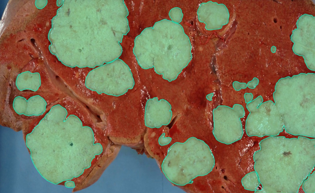
Liver Disease Analysis
Medical image annotation includes contour or polygon labeling of liver structures and specified regions in relevant medical images. These annotations help prepare structured datasets for model development, evaluation, and research involving liver imaging and related analysis tasks.

Teeth and Gum Segmentation
Dental image annotation involves identifying and labeling individual teeth and relevant oral structures using suitable annotation techniques. These labeled datasets can support the development and evaluation of computer vision models for dental image analysis.
Cancer cell Detection
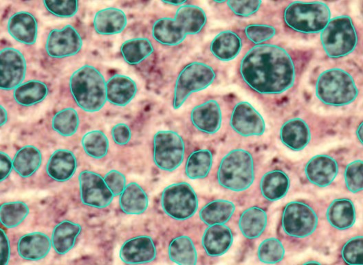
Microscopic image annotation involves labeling specified cells and regions in high-resolution microscopy images. These annotations help prepare datasets for developing and evaluating computer vision models focused on cancer cell analysis.
Intestinal parasites Detection
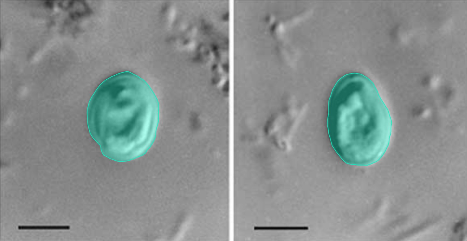
Microscopy image annotation helps identify and label visible parasites and relevant structures in supported image datasets. Contour-based annotation can be applied where appropriate to define object boundaries and prepare structured training data for image analysis models.
Lung Segmentation
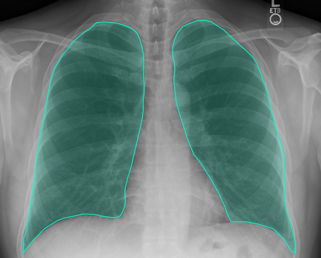
Medical image segmentation services include labeling lung regions and relevant anatomical structures in medical images. Depending on the imaging modality and project requirements, suitable segmentation techniques help create structured datasets for computer vision model training and evaluation.
Skin Biopsy
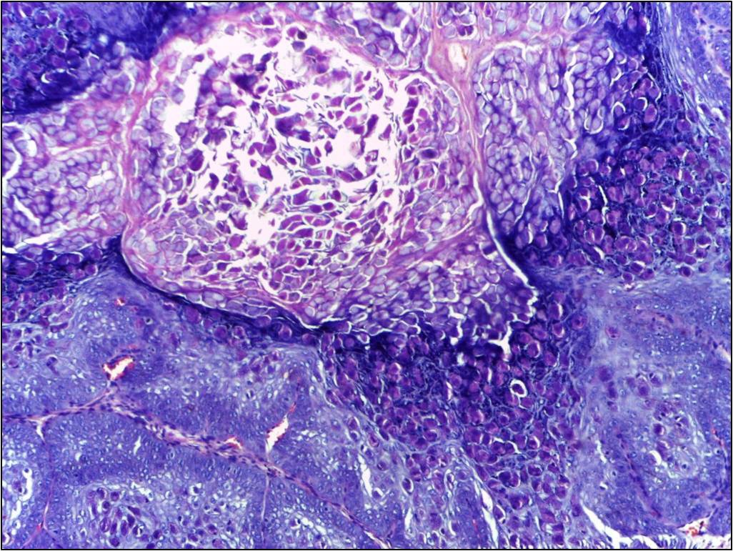
Skin biopsy image annotation involves labeling specified tissue regions and visible characteristics, including relevant structures and textures, according to project-specific guidelines. These annotated datasets can support computer vision research in histopathology and skin-related image analysis.
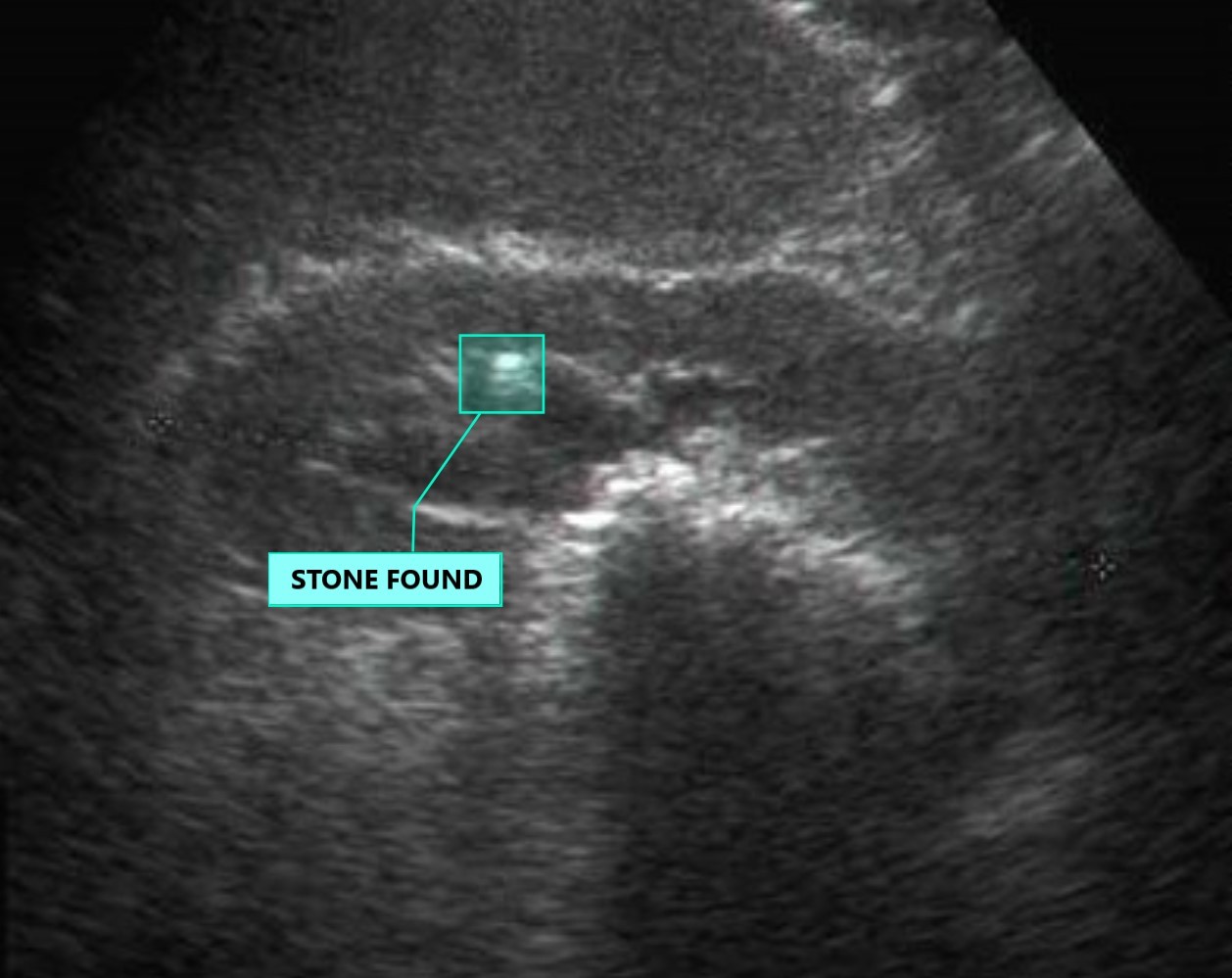
Kidney Stone
Medical image annotation helps label kidney stones and relevant anatomical regions in supported imaging datasets. These annotations can be used to prepare structured training data for computer vision models developed for kidney stone detection research and image analysis.








Frequently Asked Questions
Medical image annotation services involve labeling medical images and relevant anatomical structures to prepare datasets for training and evaluating computer vision models. Annotation tasks depend on the imaging modality, project requirements, and labeling guidelines.
Depending on the service scope, medical image annotation may cover X-rays, brain scans, retinal images, microscopic cell images, tissue samples, and other supported medical imaging datasets.
Medical image annotation is the broader process of labeling image content. Medical image segmentation specifically identifies and labels regions or structures at the pixel level, such as organs, cells, or other anatomical areas.
X-ray annotation labels specified anatomical structures or visible findings in radiographic images. These labeled datasets can support the development and evaluation of computer vision models for medical imaging research.
Annotated microscopy or radiology datasets can support research and development of models designed to identify specified abnormalities. The annotation process itself does not diagnose cancer or establish clinical accuracy.
Microscopic cell annotation involves labeling individual cells, cell boundaries, or relevant structures in microscopy images. These labels can help prepare datasets for computer vision research involving cell classification, segmentation, and analysis.


